Near-Infrared Spectroscopy
August 2014
By Kelly Shephard
The Cheatham Lab has a cool new tool for understanding nutrition and cognitive development.
It’s called a near-infrared spectroscopy (NIRS) system. NIRS offers another way to examine brain activity while people complete various tasks. Despite the fact that they look somewhat similar, NIRS is different from the electroencephalography (EEG) system we have and that many of our participants have seen before. EEG measures the electrical activity that is given off by the brain when we think. NIRS uses light to measure the flow of oxygen through the brain when we think. Oxygen moves through our bodies in hemoglobin. Oxygenated hemoglobin is what comes from our lungs and travels to our tissues in order to provide our tissues with the oxygen they need. Deoxygenated hemoglobin is what travels away from our tissues back to our lungs and heart for more oxygen. As we use our brain, oxygenated hemoglobin travels to the areas of the brain we are using and deoxygenated hemoglobin flows away from those areas. The cool thing about the hemoglobin is that light travels through oxygenated and deoxygenated hemoglobin differently. We can use this difference to pick up on the amount of oxygenated and deoxygenated hemoglobin in different areas of the brain.
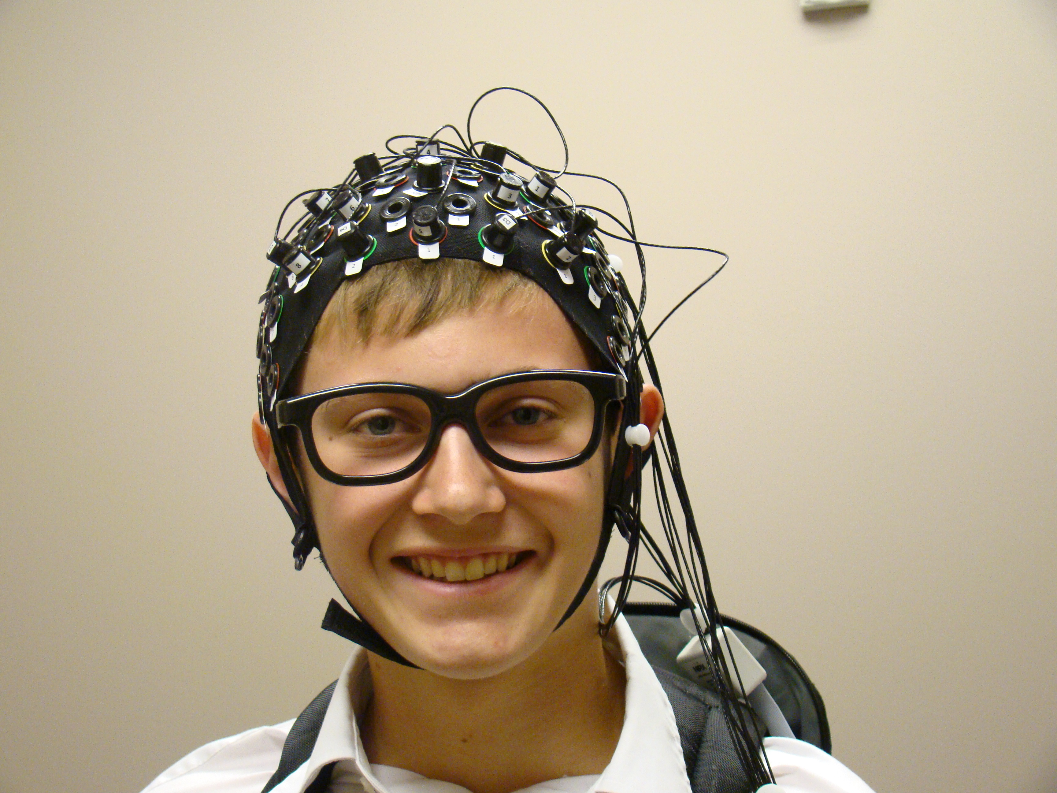 The NIRS system is made up of a cap that is kind of like a swim cap and sources (photo-transmitter probe in the diagram) and detectors (photo-receiver probe in the diagram). The sources are essentially light bulbs that give off light of very specific wavelengths. The detectors pick up on the specific wavelengths of light, and how much light the detectors pick up on tells us how much light came back after traveling through the brain tissue. We have a system with 8 sources and 8 detectors that we can put a lot of different places on the NIRS cap, which has 128 spots for sources and detectors. We can choose to measure from any area of the brain, and put the sources and detectors in the cap according to the areas of the brain we are most interested in studying.
The information about the flow of hemoglobin through the brain is useful in understanding what areas of the brain are active during different tasks. One major difference between EEG and NIRS is that EEG doesn’t give us information about where in the brain is active, it can tell us when the brain is active and how active it is. NIRS can give us a better picture of where activity is in the brain and how active it is, but it doesn’t give us very good information about when the brain is active. Using these two tools together will help us understand brain development, and how nutrition we study, like antioxidants and fatty acids, affect brain development. Right now, we are using NIRS with our study with 7- to 12-year-olds, which you can read more about in this newsletter. We can’t wait to tell you about all the cool things we can do with our brain imaging tools!